At Peel Vision, we have carefully selected our diagnostic equipment to complement the skills of our ophthalmologist to assist in the diagnosis, monitoring and treatment of a wide range of eye disease.
All tests are performed by our ophthalmic assistant, who will guide each patient through the exam to ensure that it a stress-free and comfortable experience. Our tests are painless, non-invasive, quick and safe.
Each test is performed in a seated position in front of the machine, with the chin placed on an adjustable chin-rest. The eye is focused on a target light or picture, and each eye is tested separately. Most tests take only a few seconds to a few minutes to complete.
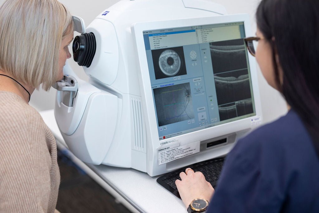
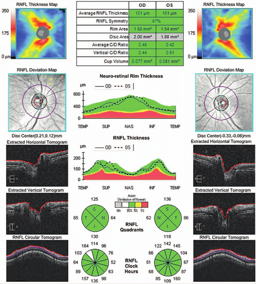
OCT scanning.
At Peel Vision, we use the Zeiss Cirrus HD-OCT 5000 scanner with Anterior Segment Premier module and Angioplex OCT-Angiography.
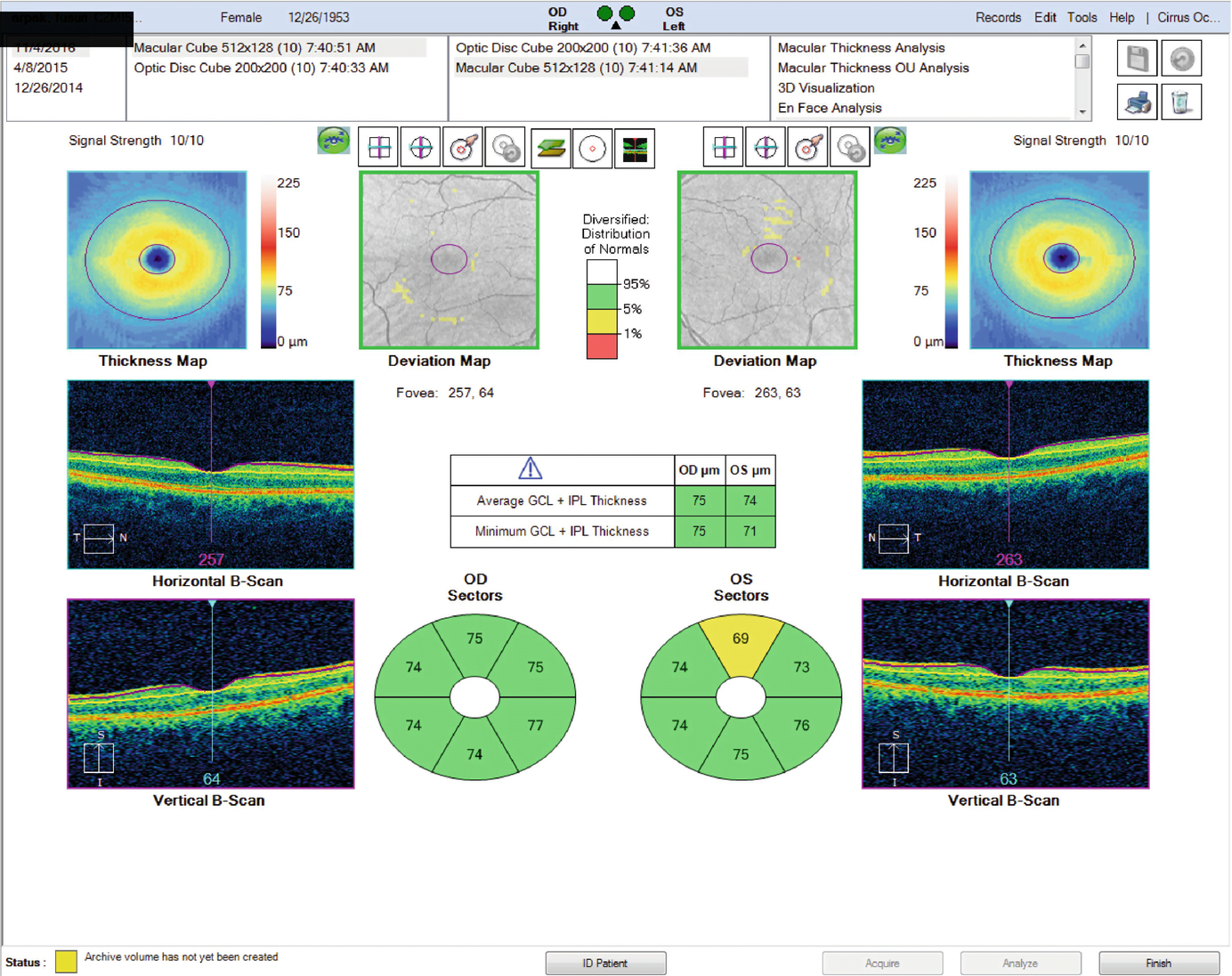
The OCT (Optical Coherence Tomography) scanner is an ultra-high resolution imaging device that takes cross-sectional images of vital structures of the front (cornea and angle) and back (retina and optic nerve) of the eye. OCT works on a principle like ultrasound to map out the layers of the back of the eye. However, instead of using sound waves, it uses near-infrared light waves. These light waves are projected and then reflected off the targeted structures of the eye. The reflected light waves are then measured and converted by a computer into high-resolution 3D colour images.
OCT scans allow the smallest of changes to be detected well before the naked eye can see it. The angioplex OCT-angiography allows further non-invasive and detailed imaging of the retinal microvasculature. OCT is a very important tool used for the diagnosis and monitoring of a wide range of sight-threatening conditions, including macular degeneration, glaucoma and diabetic retinopathy.
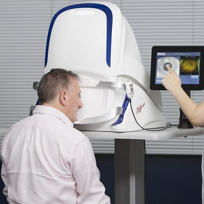
OPTOS ultra-wide-field (UWF) retinal imaging.
At Peel Vision, we use the Optos California FA device.
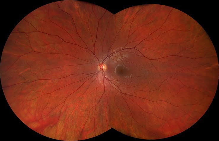
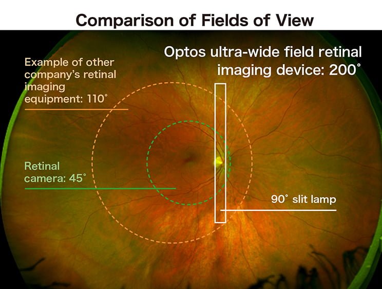
The Optos ultra-wide-field retinal imaging device is essentially a very high-resolution camera that takes detailed photographic images of the retina. Our Optos device captures more than 80% (or 200 degrees) of the retina in a single shot with unrivalled clarity and in less than ½ second. This UWF imaging allows us to see 50% more of the retina than traditional retinal photography. This means that we can discover, diagnose and treat peripheral retinal pathology earlier and more accurately.
Our Optos California FA device offers multiple imaging modalities to target specific retinal diseases, including color, red-free, choroidal and autofluorescence modes. The optional fluorescein angiography mode offers a further detailed view of the retinal microvasculature. The Optos is very useful in the diagnosis and management of retinal tears, diabetic retinopathy, macular degeneration and other vascular disorders of the retina.
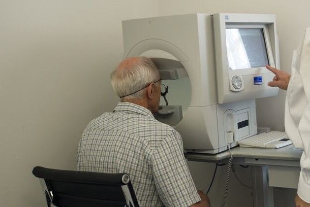
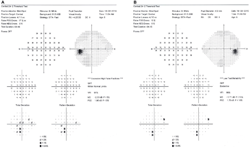
Visual field testing.
At Peel Vision, we use the Zeiss Humphrey 750i HFA-II analyser.
Visual field testing is an examination used to detect any significant loss of peripheral vision, which can occur in a variety of eye and brain diseases including glaucoma, optic neuropathy and stroke.
Most patients are not aware of any peripheral vision loss until it is very advanced. It is also difficult to detect early visual field changes with clinical or manual examination alone. Our peripheral vision is very important for many activities of daily living and for safe mobilization around our environment. A certain level of peripheral vision is required to meet Australian driving standards.
Considered the gold standard in visual field measurement, the Humphrey visual field test is a specialized automated procedure used to perform perimetry, in which the entire area of peripheral vision is mapped out whilst the eye is focused on a central target. During this test, lights of varying intensity appear in different parts of the visual field while the patient’s eye is focused on a central spot. The patient indicates whether they see the light by pushing a button. The accuracy of the patient’s perception of these lights is recorded and charted by a computer, which analyses and compares their results to a normative database to determine if their results are normal or abnormal. Each field test takes only 8-15 minutes to complete, depending on the type of field exam that is being performed.
The Humphrey Visual Field Analyzer has a very high sensitivity and specificity for detecting abnormal visual fields, as well as monitoring early changes in the field over time. Advanced software is used to chart progression of changes over time in any given patient. It is a vital tool in the management of glaucoma.
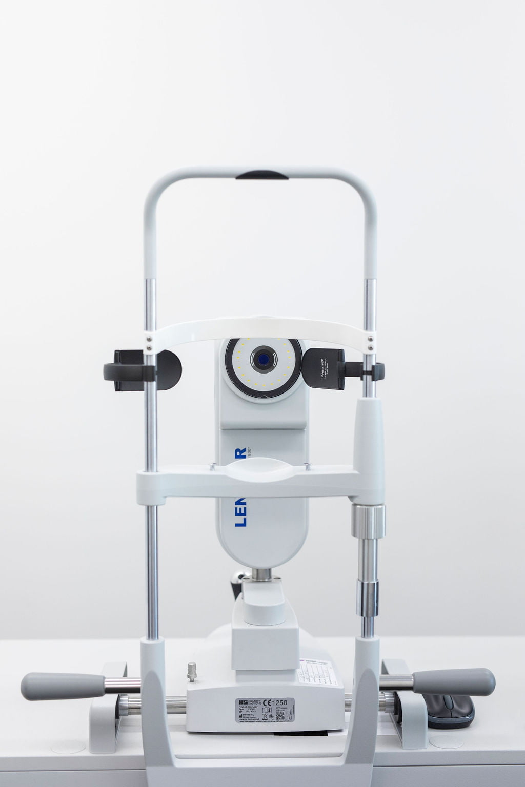
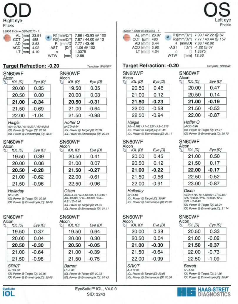
Ocular biometry.
At Peel Vision, we use the Lenstar LS-900 from Haag-Streit technologies.
Ocular biometry is the measure of the size and shape of the eye. Biometry measurements are used before cataract surgery to determine the most suitable intraocular lens implant for each patient. Precise biometry is essential to achieve the best visual outcomes in cataract surgery.
Ocular biometry uses light waves to measure various parameters of the eye and has replaced older techniques that used ultrasound (sound waves). The Lenstar is the first biometer to use the advanced technology of OLCR (Optical low Coherence Reflectometry) for all its measurements. It captures 9 vital measurements of the entire eye in 30 seconds in 1 scan. An advanced computer then analyses this data using multiple lens-calculation formulae and predictive mathematical modelling.
Dr Then will study this data to determine the most appropriate lens implant for a given eye.
The Lenstar also employs a unique and proprietary intelligent fixation system, that ensures that all measurements are taken on the patient’s visual axis, which is crucial for accuracy. If the system detects that a patient blinks or loses fixation during measurements, then scans are halted and only resumed when fixation is re-established. Often, multiple scans are done per eye to ensure the highest level of accuracy, precision and consistency is achieved.
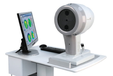
Corneal topography.
At Peel Vision, we use the Galilei G4/G6 corneal analyzer from Ziemer technologies.
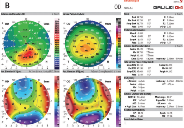
Considered a vital tool for cataract and refractive eye surgery, corneal topography takes a detailed map of the contours and thickness of the front window of the eye, known as the cornea. The Galilei analyzer takes corneal analysis to another level, being a ‘one-stop-shop’ for all the measurements required for accurate cataract and refractive surgery planning.
The Galilei analyzer is a uniquely elegant system that combines corneal curvature data (using Placido disc topography) with elevation data (using dual-Scheimpflug camera technology). This allows analysis of the curvature and thickness of both the front (anterior) and back (posterior) surfaces of the cornea, which provides more accurate data than traditional corneal topographers which only measured the anterior corneal surface.
The Galilei additionally examines disturbances in the optical function of the cornea, known as corneal aberrations, with wavefront aberrometry of both the anterior and posterior corneal surfaces. It also uses complex ray tracing to assess the total power of the cornea and axial length of the eye, which helps in calculating the parameters of a patient’s astigmatism, and the correct power and position of the intraocular lens implant required during cataract surgery.